
We talked a bit about foreign bodies last time-what common objects are, what happens when they are ingested, what to do if they are ingested, and how to prevent it from happening. For this two-part series we thought we’d give you window into how Direct Vet Services deal with foreign bodies when you bring your pet in.
When little intervention is required:Once the clinical examination is carried out, your vet will be able to inform you of what is required to treat your pet. If your pet is otherwise well and has just ingested the object presuming it is safe, we may be able to give your pet some medication to make them vomit. An object caught in the mouth, for example a bone caught on a tooth or a string around the tongue, may be easily removed in a consult or under sedation or light anaesthesia. Other times if the object is small enough and is of no danger to allow it to continue, it is possible to monitor your dog’s appetite, clinical signs and stools to ensure the foreign object has passed through safely. We have retrieved a few engagement rings this way!
Testing:
Your pet may require a blood test to rule out other causes of the clinical signs as well as checking for electrolyte imbalances, degree of dehydration, and other problems related to foreign bodies. If we need to put your pet under anaesthetic,we need to run tests to ensure that other organ systems have not been compromised which increases the risk of further complications. We may choose to start with Intravenous fluids will help to rehydrate your pet and rebalance electrolytes, especially potassium, which is imperative for heart muscle function.
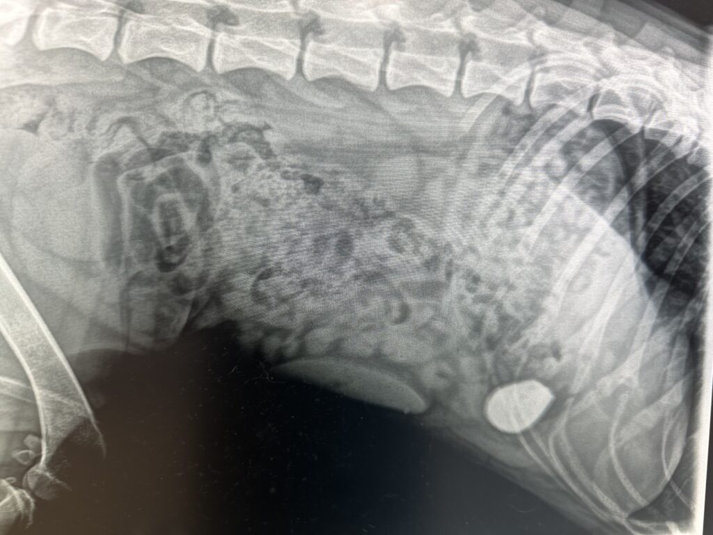
X-rays:
We may recommend X-rays to get a better idea of what the foreign body is or where in the gut it is located. We will look for the foreign body as well as changes in the gas pattern within the intestines (suggestive of a blockage), thickening or ruffling of the intestinal wall, and evidence of motility. A foreign body isn’t always obvious or visible on an X-ray however.Other studies:Sometimes foreign bodies can be hard to find or are difficult to see via an X-ray.Dish sponges and fabric are a classic example of hard-to-find foreign bodies and sometimes a contrast agent or barium study is recommended as this can outline a foreign body or highlight a blockage. An abdominal ultrasound can also be useful in assessing the abdomen and guide treatment. Remember though these are all diagnostic tools, we then must fix the problem.
Treatment:
Scope or induced vomiting:
Scope or inducing vomiting may be possible if the object is in the stomach and suitable for removal in this way. The best ever case where we induced vomiting was a pug that swallowed a green squishy frog toy. When we made the pup vomit,the toy frog quite literally looked like it leaped from the dog’s mouth, much to the amusement of the staff. Once the object has passed the stomach, scope and vomiting are not useful in most cases and it’s off to surgery we go. In the large intestine, unless there is an obvious problem we may choose to wait. Being wider than the small intestine, once foreign bodies have made it this far,they usually keep going.

Surgery:
If the object is not suitable to be removed via scope or induced vomiting, then we need to perform a laparotomy and remove it. A foreign body located in the oesophagus may be diagnosed and possibly removed via a flexible endoscope (a small tubular camera placed down the oesophagus). We do not use vomiting to retrieve objects in the oesophagus as they are “caught” and would risk perforation.The most common objects that lodge in this location are bone shards and fish hooks although we did have a pen once
Prompt surgery may be recommended to remove the foreign body from the intestines to prevent blockage and potentially serious consequences. The longer the foreign body is present the more problematic is the outcome in most cases,although I did have a Doberman once that ate the rubber head of a spatula, the owners thought he must have passed it as other than the very odd vomit he never showed any signs. Then 8 months later it became stuck in the pyloris, ( the muscular opening between the stomach and small intestine) and that was it.Sometimes a section of the bowel may need to be removed if it is perforated or badly damaged and likely to break down post-surgery. A ruptured gut and resulting peritonitis means a much poorer prognosis as the animal is often septicaemic at this stage and multiple other organs have been damaged by the toxins.
If in doubt!
If you suspect or have seen your pet eat a foreign body of any kind, or if you have noticed your dog is showing any of the clinical signs, it is important that you contact your vet and remember time might be of the essence.If you have any concerns or further questions about your pet please book in for a consultation online or by calling our awesome reception team on (03) 9369 1822.